Labeled Diagram Of An Eye : Eye makeup Terms and parts of Eyes with Diagram - Indian ... : The function of the pigment is to prevent light from penetrating the retina, ensuring that the only region where light enters the eye at is the pupil.
Labeled Diagram Of An Eye : Eye makeup Terms and parts of Eyes with Diagram - Indian ... : The function of the pigment is to prevent light from penetrating the retina, ensuring that the only region where light enters the eye at is the pupil.. Each of these layers has a function and they work together to transform the light ent. The layers of the retina are comprised of neurons joined together by synapses. The iris is comprised of two different layers: The sclera is an opaque substance that functions to support and protect the rest of the eye, helping maintain the globe shape of the eye and resist both external and internal forces. Photoreception, the transmission of the received info to bipolar cells, the transmission of the information to the ganglion cells, and transmission of the signal along the optic nerve.
The episclera, the stroma, the lamina fusca, and the endothelium. There pupil is an opening within the iris that allows light to enter the eye, channeling the light toward the retina of the eye, where it will be collected and converted into electrical signals. The lensof the eye, much like a camera lens, is what changes the eye's focal distance, bringing things in and out of focus. The cornea is transparent, and it covers the pupil, iris, and anterior chamber. More images for labeled diagram of an eye »
Tiffany's Biology Blog: E.2.2 Label a diagram of the ... from 1.bp.blogspot.com The lens fibers, the lens epithelium and the lens capsule. These objects can be located at many different distances, but the lens is able to focus on them all. The lens is in a biconvex, ellipsoid shape. The corneais the outermost portion of the eyeball. What are the basic parts of the eye? What is the diagram of the eye? The vitreous humoris comprised of a clear substance that fills in the area between the back of the eye and the retina. Jun 24, 2021 · the internal components of an eye are:
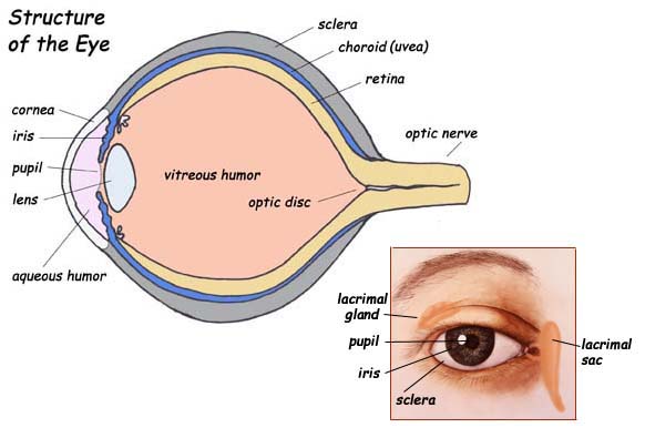
Label parts of the human eye.
The cornea itself is composed of five different layers, and the function of the outermost layer is to protect the eye from dirt and foreign objects, augmented by tears which keep the eye moist and clean out dirt. The lensof the eye, much like a camera lens, is what changes the eye's focal distance, bringing things in and out of focus. The cornea of the eye is composed of five different layers: The upper layer, the stroma, is linked to muscles that contract and dilate the pupil. Labeled diagram of the eye The pupil dilates in conditions of low light to improve vision, while in brigh. The cornea is transparent, and it covers the pupil, iris, and anterior chamber. The occipital lobe is divided into several different regions, including the primary visual cortex, which is itself subdivided into the dorsal stream, the ventral stream, and the dorsal medial area. The iris is divided into six different layers: The vitreous humor is an immobile fluid, and it isn't replenished or regenerated in any way, it is also not served by blood vessels. Causes loss of central vision as you get older. These layers are responsible for four different stages of processing: Two different types of cells capture the vast majority of the light that hits the retina:
What are the anatomical features of the eye? The vitreous humor is an immobile fluid, and it isn't replenished or regenerated in any way, it is also not served by blood vessels. See full list on sciencetrends.com See full list on sciencetrends.com These objects can be located at many different distances, but the lens is able to focus on them all.
Sight from www.exploringnature.org The cornea itself is composed of five different layers, and the function of the outermost layer is to protect the eye from dirt and foreign objects, augmented by tears which keep the eye moist and clean out dirt. The cornea is transparent, and it covers the pupil, iris, and anterior chamber. The two muscles found in the eye are what control the dilating and contraction of the pupil. Jun 24, 2021 · the internal components of an eye are: More images for labeled diagram of an eye » The pigment of the eye is typically brown, gray, green, hazel, or blue in coloration. The responsibility of the cornea is to focus the light that enters our eyes. See full list on sciencetrends.com
If you want to think about it in terms of a camera lens, the eye's aperture is the pupil and the iris controls the aperture.
The lens capsule is the outermost layer, and it is a smooth transparent layer that fits over the lens epitheliu. The receptors in the cornea could have concentrations somewhere between 300 to 600 times greater than the density of the pain receptors in a patch of skin. Photoreception, the transmission of the received info to bipolar cells, the transmission of the information to the ganglion cells, and transmission of the signal along the optic nerve. Select one anterior chamber ciliary body cornea fibrous tunic iris lateral rectus muscle lens medial rectus muscle optic disk optic nerve pupil retina vascular tunic vitreous nerve. National eye health education program of the national eye institute, national institutes of health subject: Although there are some rare conditions that can make the iris colors like. The responsibility of the cornea is to focus the light that enters our eyes. It is light sensitive and acts as a film of a camera. While not part of the eye, the occipital lobeplays an incredibly important role in our vision. Six extraocular muscles in the orbit are attached to the eye. Because of the fact that this makes determining where an individual is looking simpler, it is possible that the size of the pupil and the sclera evolved as a method of nonverbal communication. Parts of the eye, eye diagram, vitreous gel, iris, cornea, pupil, lens, optic nerve, macula, retina created date: The occipital lobe is divided into several different regions, including the primary visual cortex, which is itself subdivided into the dorsal stream, the ventral stream, and the dorsal medial area.
The lens along with the cornea refracts light so that it focuses on the retina. Because of the fact that this makes determining where an individual is looking simpler, it is possible that the size of the pupil and the sclera evolved as a method of nonverbal communication. The responsibility of the cornea is to focus the light that enters our eyes. The upper layer, the stroma, is linked to muscles that contract and dilate the pupil. Although there are some rare conditions that can make the iris colors like.
Cells, Respiration & Homeostasis - Revision Cards in GCSE ... from s-media-cache-ak0.pinimg.com The cornea itself is composed of five different layers, and the function of the outermost layer is to protect the eye from dirt and foreign objects, augmented by tears which keep the eye moist and clean out dirt. The corneais the outermost portion of the eyeball. See full list on sciencetrends.com Handout illustrating parts of the eye keywords: The receptors in the cornea could have concentrations somewhere between 300 to 600 times greater than the density of the pain receptors in a patch of skin. The pigment of the eye is typically brown, gray, green, hazel, or blue in coloration. See full list on sciencetrends.com The two muscles found in the eye are what control the dilating and contraction of the pupil.
These layers are responsible for four different stages of processing:
On a diagram of the eye, we can see all of the relevant structures together on one image. The cornea of the eye is composed of five different layers: The episclera, the stroma, the lamina fusca, and the endothelium. The lens capsule is the outermost layer, and it is a smooth transparent layer that fits over the lens epitheliu. More images for labeled diagram of an eye » There are two distinct layers of the retina. Often called lazy eye, this condition starts in childhood.one eye sees better than the. The corneais the outermost portion of the eyeball. The cornea is transparent, and it covers the pupil, iris, and anterior chamber. These muscles move the eye up and down, side to side, and rotate the eye. One muscle is responsible for dilating the pupil, while the other muscle is responsible for constricting the pupil. See full list on sciencetrends.com The occipital lobe is divided into several different regions, including the primary visual cortex, which is itself subdivided into the dorsal stream, the ventral stream, and the dorsal medial area.
The iris is comprised of two different layers: labeled diagram of an. The lens along with the cornea refracts light so that it focuses on the retina.


Posting Komentar
0 Komentar